STED workshop 06. - 09. December 2016
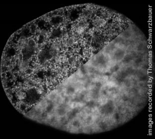 We are pleased to announce a STED course co-organized by Leica Microsystems and the Core Facility Bioimaging. The event is dedicated to the use of STED superresolution microscopy for fixed and living, thin and thick samples, deconvolution and analysis of results.
The target audience are users who began to use STED or will begin soon. The basics of laser scanning microscopy (e.g. confocal microscopy) should already be familiar to the participants.
We will accept up to 18 participants. For the six hands-on sessions they will be divided into three groups, two of which will work on Leica STED microscopes while the third will cover related topics (see below for details).
 Time and Location
Start: 06. December, 13:00
End: 09. December, 13:00
Core Facility Bioimaging
Biomedical Center (BMC)
Ludwig-Maximilians-Universität München
Großhaderner Str. 9
82152 Planegg-Martinsried, Germany
The BMC is a few meters outside the city limits of Munich. See below for travel information.
Leica Microsystems kindly provided a flyer for the course: download.
Preliminary program
Day 1: December 6, 2016 (Tuesday)
13:00 Welcome (Steffen Dietzel and Nathalie Garin)
13:15 STED principle (Patrick Zessin)
13:45 Fixed sample preparation and fluors for STED (Jana Dohner)
14:15 Open discussion: How do you fix, which fluors do you use (Chair: Jana Dohner / Nathalie Garin)
14:45 Coffee break
15:15 Live cell imaging with STED (Ulf Schwarz)
16:00 Hands-on 0: How to use the software. Split in two groups
17:15 Short presentation of the participants, Part 1 (5 min / person, 12 of 18)
19:00 End of day 1
Day 2: December 7, 2016 (Wednesday)
 09:00 Comparison of DNA labels for STED microscopy (Steffen Dietzel, Andreas Thomae)
09:30 Deconvolution: Concepts and applications (Hans van der Voort)
10:15 Coffee break
10:45 The most elaborate is not always the best: 5 colors with up to 140 nm resolution with confocal deconvolution / HyVolution (Patrick Zessin)
11:15 Application examples (Nathalie Garin)
11:45 How to check your systems performance (Ulf Schwarz)
12:15 Lunch
13:15 Short presentation of the participants, Part 2 (5 min / person, 6 of 18)
14:45 Hands-on 1
16:45 Coffee break
17:15 Hands-on 2
19:15 End of day
Day 3: December 8, 2016 (Thursday)
09:00 Hands on 3
11:00 Coffee break
11:30 Hands-on 4
13:30 Lunch
14:30 STED microscopy of the living mouse brain (Katrin Willig)
15:30 Coffee break
16:00 Hands-on 5
18:00 End of day
Day 4: December 9, 2016 (Friday)
09:00 Hands-on 6
11:15 Feedback and goodbye
12:00 End of course, lunch
Hands-on sessions
- STED with fixed samples I: Cytoskeleton stainings, gold beads and others.
- STED with fixed samples II: Thick samples, nanorulers
- STED with Fixed samples III: continued from above, user samples
- Deconvolution and Quantification of STED images
- STED with living cells
- Hyvolution as a 'more colors alternative' to STED: Confocal microscopy with small pinhole, sensitive detectors and deconvolution
Teachers and Organizers
Dr. Steffen Dietzel, Head of the Core Facility Bioimaging
Dr. Jana Döhner, Center for Microscopy and Image Analysis, University of Zurich
Dr. Nathalie Garin, Leica Microsystems
Ulf Schwarz, Leica Microsystems
Dr. Remco T.A. Megens, Institut für Prophylaxe und Epidemiologie der Kreislaufkrankheiten, Klinikum der Universität München
Dr. Hans van der Voort, Scientific Volume Imaging, Netherlands
Dr. Katrin Willig, Center for Nanoscale Microscopy and Molecular Physiology of the Brain, University Medical Center and Max Planck Institute of Experimental Medicine, Göttingen, Germany
Dr. Patrick Zessin, Leica Microsystems
Registration
The registration fee is 400 Euro. It does NOT include accomodation.
Registration deadline is Monday, 24. October. We appreciate earlier registrations. Please go to the registration page to register. We will select the participants and inform all applicants as soon as possible after the registration deadline. Registration will be complete only after we receive the fee. Payment details will be provided after the selection process.
Travel and accomodation
The Biomedical Center (BMC) is located at the south-western edge of Munich, on the Großhadern-Martinsried campus. The campus web site has a page with detailed instructions how to get here.
We have made a pre-reservation with a nearby hotel (~ 15 min walk) which the selected paricipants can use in their own name or ignore, as they see fit. This reservation would be available from 05.12. - 09.12.2016 for 89 Euros per room per night (incl. breakfast).
Supporters
Apart from our coorganizer Leica Microsystems the following organizations support us by providing personell or material for this workshop:
 Institut für Prophylaxe und Epidemiologie der Kreislaufkrankheiten, Poliklinik, Klinikum der Universität München, Ludwig-Maximilians-Universität München
 Scientific Volume Imaging (SVI), manufacturer of the deconvolution software Huygens
 Gattaquant, producer of nanorulers and 'DNA beads' to measure performance of a microscope
 Spirochrome, producer of silicon rhodamine (SiR) dyes.
Downloads
- STED-Workshop December 6-9 2016 (258 KByte)
- van der Voort (181 KByte)
- Willig (126 KByte)


