Materials for Teaching
This page contains material for download which can be used for teaching courses on light microscopy. It may have limited use for self-study since a certain level of prior knowledge is expected for its application
Presentations
The Powerpoint presentations follow the suggestions of the Workgroup 5 'Training of facility users' of the German Bioimaging (GerBi) initiative on how to structure light microscopy courses. The presentations are, however, not complete lectures on the respective course units, copyrighted material was removed. The material provided is under a creative commons license which allows free distribution. License details are given in the files.
Materials for the following units are provided:
1 Basic Microscope Optics (BMO)
2 Basics of Digital Imaging (BDI)
4 Image processing and presentation (IPP)
6 Basic Laser Scanning Confocal Microscopy (this is a zip file which contains the presentation and two movies)
Alternative download site: These presentations plus some materials provided by others are also available from the German BioImaging Website. Click on the 'Material'-link at the end of each unit here.
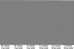
Template for diffraction patterns
 This template is for the production of photographic slides (diapositives) which can be used to generate diffraction patterns with a laser beam. Six columns represent six patterns with lines, each pattern with lines twice as thick as the one left to it.
When this 8192 × 5464 Pixel image is used as source for a slide production with 8192 lines, the number of line pairs (lp, one pair is one black and one white line) per millimeter will be approximately what is written at the bottom of each column. The production process is far from perfect. Microscopic inspection of such slides showed that the 60 lp/mm pattern was very badly resolved during the slide production process. Accordingly, the diffraction pattern generated by this area of the slide was very weak (but just recognizable). From the production of a different pattern we found that a 1 px wide line pattern resulted in a gray area. For the larger patterns, we noted that the white lines in the slide turned out to be thicker than the black ones.
We ordered our slides from http://www.diabelichtung.info. The mailing came from WightSlide, Cowes, UK, so these two companies either work together or are the same. Mentioning this particular company is solely for informational purposes. We have no reason to believe that other services could not provide similar or possibly even better quality.
Download file 'StreifendiaText.png' from this server (right-click and 'save target as')
Alternative download site: page at the German BioImaging (GerBi) web site
File created by Anna Klemm and Steffen Dietzel, Core Facility Bioimaging at the Biomedical Center of the Ludwig-Maximilians-Universität München. Thanks to Jan Peychl, Dresden, for inspiration and to Peter Evennett from the Royal Microscopical Society who provided Jan with slides with stripe patterns which are thus direct ancestors of the image provided here.
Downloads
- SD_1_Basic Microscope Optics (BMO) (4 MByte)
- SD_2_Basics of Digital Imaging (BDI) (3 MByte)
- SD_4_Image processing and presentation (IPP) (29 MByte)
- SD 6 confocal (32 MByte)


