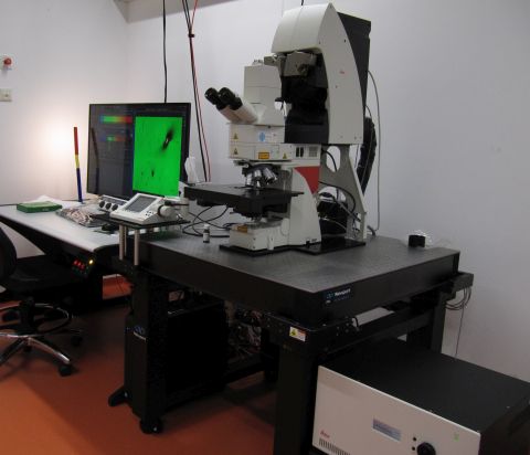
Malpighi: Upright confocal with physiology stand
 Named after Marcello Malpighi (1628 – 1694), an Italian biologist and physician who, among other things, discovered the blood capillaries (Wikipedia de|en).
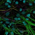
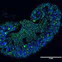
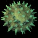
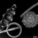
Gallery
 |
 |
 |
 |
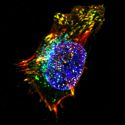 |
For more information and for high resolution images please go to the gallery subpage.
Summary
This Leica SP8X WLL upright confocal microscope has a DM6 stand. 405 nm and pulsed "white light" Laser (470 - 670 nm) for excitation, gated hybrid detectors and PMTs for detection.
This microscopy is perfect for confocal microscopy for
- samples that require an upright microscope.
- any kind of fixed samples. (Less trouble with immersion oil compared to inverted microscope).
- living (cultured) cells that should be observed in a dish with a dipping objective.
This microscope is not perfect for
- high resolution or sterile observation of living (cultured) cells. Use Kellner instead, an inverted confocal.
- FRAP experiments that need strong laser power: the 1.5 mW of several wavelengths of the white light laser may be combined, but that still may not suffice. If you can use an inverted confocal for FRAP use Kellner. If you need an upright confocal for FRAP, try Hooke. Apart from the multi-photon laser Hooke has 488 nm (20 mW), 552 nm (20 mW) and 638 nm (30 mW) lasers.
Optics
Condenser
P 0.9 S1. The 0.9 indicates the numerical aperture.
Objectives
This is the standard configuration of the objective turret:
- HCPL FLUOTAR 10x/0.30, working distance (WD) 11.0 mm. Usable with and without coverslip.
- HC PL APO 20x/0.75 CS2. WD 0.62 mm. Must be used with 0.17 mm coverslips. No immersion.
- HC PL APO 20x/0.75 IMM CORR CS2. Usable with and without coverslip, with water, glycerol or oil. WD depends on immersion, e.g. 0.67 mm for water with coverslip.
- HC PL APO 40x/1.30 Oil CS2. WD 0.24 mm.
- HC PL APO 63x/1.30 GLYC CORR CS2. For glycerol immersion (ne=1.451 @ 21°C). WD 0.3 mm. Color corrected for 21°-37°C. Correction collar for temperature, coverslip thickness or refractive index (80% glycerol/ 20% water).
- Obj. HC PL APO 63x/1.40 OIL CS2. WD 0.14 mm.
DIC Prisms
Any laser line can be used to create DIC images with the transmission detector. This option can be selected in the software. The correct prisms for the respective objective will then be selected automatically if available. Caution: a DIC prism in the beam path has a negative impact on your confocal (fluorescence) resolution!
Conventional fluorescence
Meant for visual inspection only, not for documentation. Excitation with SoLa white light LED. Filters for blue (exc. 385-335, dichr. 455, em. LP 450-490), green (exc. 450-490, em. LP 515) and orange (exc. 515-560, em. LP 590) are available.
Confocal Light Path
Excitation Lasers
Two lasers are available. The 405 nm laser (continuous wave) is usually used for Dapi or related dyes. It has an output of 50 mW which can be attenuated with an AOTF.
The WLL2 "white light laser" emits all wavelengths between 470 and 670 nm. Of those, up to 8 can be selected simultaneously. Each selected wavelength has about 1.5 mW power which can be attenuated with the AOTF. The WLL2 is a pulsed laser (80 MHz), allowing time gating of fluorescence life times in combination with HyD detectors. An article on the Leica Science Lab Website describes this laser in further detail.
Beam Splitter
Instead of dichroic filters, an optical crystal is used as beam splitter, the acousto-optical beam splitter (AOBS). You don't need to know how it works, but if you want to, there is an article on the Leica Science Lab. Depending on the selected excitation wavelengths, the settings for this filters are made automatically.
Detectors
The system contains one transmission detector with which a non-confocal image can be recorded, bright field or DIC. (Caution: a DIC prism in the beam path has a negative impact on your confocal (fluorescence) resolution!).
The detectors for the confocal images are all in the spectral scan head which allows continuous detection between 400 and 800 nm. We have 2 Photomultiplier Tubes (PMTs; Hamamatsu R 9624)) and 3 hybrid photo detectors (HyDs). Caution: The HyDs are more sensitive (45% QE at 530 nm) but also more easily damaged! As explained during hands-on training, if you have an untested sample we highly recommend to start with the PMTs to get an impression of signal intensity. See "Sensors for True Confocal Scanning" (Leica Science Lab) for more infos on HyDs.
Scanners
Two different scan modes are available. The "normal" scanner is also called Field of View (FOV) scanner. It has a very large maximal field of view with up to 8192x8192 pixels and speeds between 1-1800 lines per second (Hz). With 512x512 pixels it can record up to 7 frames per second. Zoom factor can be 0.75 - 48x. In 09/2020 the scanner electronics was upgraded. It now allows linear scanning (previously sigmodal) and vastly improved light collection. The 'duty cycle' indicates the percentage of time that is actually used for photon collection. In unidirectional (normal) scan mode it went up from 25% to 65%, in bidirectional scan mode from 50 to 75%.
The 8 kHz resonant scanner does a fixed number of 8000 lines per second, or 16000 in bidirectional mode. It records up to 25.72 frames at 512x512 pixels (pixel dwell time 72 ns). Maximal image size is 2496x2496 pixels (6.13 frames/s, pixel dwell time 15 ns). Zoom factor 1.25x - 48x.
Stage
The original Scientifica MMBP stage was replaced in 09/2020 with a "CFS scanning stage" which is more convenient for slides and other standard samples. A Scientifica stage is available at Gaviola. The CFS stage holds standard stage inserts (116 x 160 mm) and can move over 130 x 73 mm. Maximum speed: 60 mm/s. The stage does not move in z, but the objective is moved up and down to make z-stacks.
For your Methods Section
If you used this microscope in your work, here is a suggestion for your methods section. You are welcome to copy it, but make sure to adapt it to the objectives, pixel size, dyes, filter settings etc. that you actually used.
"Confocal microscopy was performed at the core facility bioimaging of the Biomedical Center with a Leica SP8X WLL microscope, equipped with 405 nm laser, WLL2 laser (470 - 670 nm) and acusto-optical beam splitter. Images were acquired with a 63x1.4 objective, image pixel size was 80 nm. The following fluorescence settings were used: Dapi (excitation 405; emission 410-470), GFP (489; 492-550), Cy3 (558; 560-600) and Cy5 (650; 652-700). Recording was sequentially to avoid bleed-through. GFP and Cy3 were recorded with hybrid photo detectors (HyDs), Dapi and Cy5 with conventional photomultiplier tubes."
You are very welcome to ask the facility staff for help to get your microscopy section right. Acknowledgement of the core facility is extremely important for us justify past funding and thus to secure future funding, so that we will be able to continue to provide good services to you.