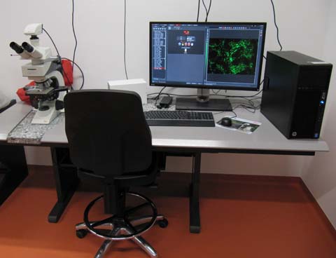
Jabłoński: Fluorescence microscope with CCD-Camera
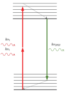 Named after Aleksander Jabłoński (1898 - 1980), a Polish physicist who developed the Jablonski diagram to explain fluorescence (Wikipedia de|en).
Summary
Leica DM2500 upright fluorescence microscope with five color channels and with a sensitive black and white camera.
Optics
Condenser achr. apl. A 0.9 (P), CC.
Objectives
HC PL FLUOTAR 5x/0.15. Working distance (WD) 13.7 mm. Usable with or without coverslip.
HC PL FL 10X/0.30. WD 11.0 mm. Usable with or without coverslip.
HC PL FLUOTAR 20x/0.50. WD 1.15 mm. 0.17 mm coverslip must be used.
HC PL FLUOTAR 40X/0.8. WD 0.40 mm. 0.17 mm coverslip must be used.
HC PL APO 63x/1.40-0.60. WD ??. 0.17 mm coverslip must be used.
HCX PL APO 100x/1.40-0.70 Oil. WD 0.09 mm. 0.17 mm coverslip must be used.
The 63x and 100x objectives have an adjustment ring to reduce the numerical aperture from 1.4 down to 0.6 or 0.7, respectively. Such a reduction makes sense only in rare cases. Usually, the ring should be set to the maximum position. That is the position were the excitation light passed through the objective is most intense.
Fluorescence
Light source
Lumencor SOLA-SM-II (365nm version) white LED.
Filters
Filter system DAPI ET: Excitation BP 325-375; dichroic LP 400; emission BP 435-485.
Filter system L5 ET: BP 460-500; LP 505, BP 512-542. (Green fluorochromes: FITC, Alexa 488, GFP)
Filter system Cy3: BP 542-568; LP 575; BP 579-631. (Orange fluorochromes: TRITC, Cy3)
Filter sytem TXR: 540-580; 585; 592-668 (Near red fluorochromes: Cy3.5, Texas Red, Alexa 594, mCherry).
Filter system Y5 ET: 590-650; LP660; 662-738. (Deep Red fluorochromes: Cy5, SiR)
All values in nm.
Camera
Leica DFC3000 G with Sony ICX455 interline CCD chip with micro lenses. 1296 x 966 pixels (1.3 Mpixel). Chip pixel size 3.75 µm. The camera is mounted with a 0.4x C-mount adapter, effective pixel size is thus 9.375 µm. Example: With the 10x objective, image pixel size is 0.9375 µm. See table for other objectives. Image pixel size is an important parameter since, together with the NA of the objective, it allows to calculate the actual resolution of the image
Leica Website on the DFC3000 G. Brochure download: pdf (4 Mb)
| Objective | c-mount | Image Pixel Size |
|---|---|---|
| 5x | 0.4x | 1.875 µm |
| 10x | 0.4x | 0.935 µm = 935 nm |
| 20x | 0.4x | 0.469 µm = 469 nm |
| 40x | 0.4x | 0.234 µm = 234 nm |
| 63x | 0.4x | 0.149 µm = 149 nm |
| 100x | 0.4x | 0.0935 µm = 93.5 nm |
For your Methods Section
If you used this microscope in your work, here is a suggestion for you method section. You are welcome to copy it, but make sure to adapt it to the objectives, dyes and filters that you actually used.
"Fluorescence microscopy was performed at the core facility bioimaging of the Biomedical Center with a Leica DM 2500 microscope, equipped with a DFC3000 G CCD camera. Images were acquired with a 100x1.4 objective, image pixel size was 94 nm. The following fluorescence channels were used: Dapi (exc 350/50; em 460/50), GFP (480/40; 527/30), Cy3 (546/10; 585/40) and Cy5 (620/60;700/75m)."
You are very welcome to ask the facility staff for help to get you microscopy section right. Acknowledgement of the core facility is extremely important for us to secure future funding and thus to provide good services to you.