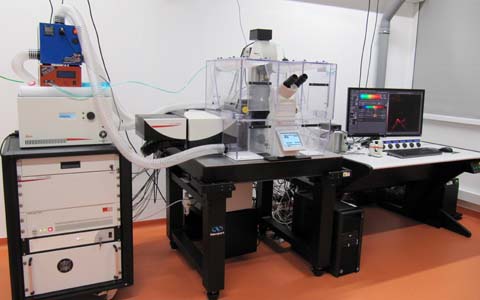
Kellner: Inverted confocal with STED and light sheet
 Named after Carl Kellner (1826 - 1855), German microscope builder and entrepreneur (Wikipedia de|en). Manufacturer's designation: Leica SP8X STED 3D DLS. Inverted confocal microscope with 405 nm and pulsed "white light" Laser (470 - 670 nm). STED depletion lasers at 592, 660 and 770, the later one pulsed, for depletion in 2D and 3D. Fluorescence lifteime imaging (FLIM). An add-on allows light sheet microscopy.
Summary
This instrument is an inverted Leica SP8X WLL confocal microscope with STED and digitial light sheet extensions. Based on a DMi8 stand it is equiped with 405 nm, a pulsed "white light Laser" (WLL; 470 - 670 nm) and a powerful Argon laser for excitation or bleaching, gated hybrid detectors and PMTs for detection. The WLL can be used for Fluorescence Lifetime Imaging (Leica FALCON), also in combination with STED (Tau-STED). An incubation box allows temperature and CO2 control.
This microscope can be used for
- high resolution and confocal microscopy of living (cultured) cells.
- FRAP experiments on such cells with the high power argon laser.
- FLIM
- super resolution microscopy with the STED module, also in combination with FLIM to increase signal to noise ratio (Tau-STED).
- light sheet microscopy.
This microscope should not usually be used for normal confocal microscopy of fixed samples. Use Malpighi instead, an upright mircoscope with similiar confocal equipment (Less trouble with immersion oil compared to inverted microscope).
Optics
Condenser
S28/0.55. The 0.55 indicates the numerical aperture.
Objectives for confocal and STED microscopy
Not all of the following objectives can be mounted at the same time. Please inquire if you are not sure that the objective you need is actually screwed into the turret. The core facility staff will be happy to provide the required objective for you.
- HCPL FLUOTAR 10x/0.30, working distance (WD) 11.0 mm.
- HC FLUOTAR L 25x/0.95 W VISIR 0.17. Must be used with 0.17 mm coverslips and immersion water. WD 2.5 mm
- HC PL APO 63x/1.20 W motCORR CS2. Water immersion with motorized corretion collar for coverglases 0.14 - 0.18. WD 0.3 mm. An automated water dispenser is available.
- HC PL APO 20x/0.75 IMM CORR CS2. Multi-immersion objective (water, glycerol, oil; adjust correction collar to your immersion medium!) for use with or without coverslip. WD 0.67 mm if used with water and 0.17 mm coverslip.
- HC PL APO 40x/1.30 Oil CS2. WD 0.24 mm.
- HC PL APO 93X/1.30 GLYC motCORR – STED WHITE. Motorized correction collar allows adjustment of refractive index mismatch, e.g. to image deeper in a sample or for various mounting media.
- 100x/1.40 OIL STED White objective. This objective has a particularly low chromatic aberration in z-direction <50 nm to ensure overlay of excitation focus and depletion donut in STED application. See image here.
DIC Prisms
Any laser line can be used to create DIC images with the transmission detector. This option can be selected in the software. The correct prisms for the respective objective will then be selected automatically if available. Caution: a DIC prism in the beam path has a negative impact on your confocal (fluorescence) resolution!
Conventional fluorescence
Meant for visual inspection only, not for documentation. Excitation with SoLa white light LED. Filters for blue (LED 405), green (exc. 450-490, em. LP 515) and orange (exc. 515-560, em. LP 590) are available.
Confocal Light Path
Excitation Lasers
Three lasers are available. The 405 nm laser (continous wave) is usually used for Dapi or related dyes. It has an output of 50 mW which can be attenuated with an AOTF.
The WLL2 "white light laser" emits all wavelengths between 470 and 670 nm. Of those, up to 8 can be selected simultaneously. Each selected wavelength has about 1.5 mW power which can be attenuated with the AOTF. The WLL2 is a pulsed laser (80 MHz), allowing time gating of fluorescence life times in combination with HyD detectors. An article on the Leica Science Lab Website describes this laser in further detail. For FLIM, a pulse picker allows to select 40 or 20 MHz to allow measuring long life times.
An additional powerfull Argon laser is available for bleaching experiments like FRAP with the following excitation lines: 458 nm, 476 nm, 488 nm, 496 nm, 514 nm.
Beam Splitter
Instead of dichroic filters, an optical crystal is used as beam splitter, the acousto-optical beam splitter (AOBS). You don't need to know how it works, but if you want to, there is an article on the Leica Science Lab. Depending on the selected excitation wavelengths, the settings for this filters are made automatically.
Detectors for confocal and STED microscopy
The system contains one transmission detector with which a non-confocal image can be recorded, bright field or DIC. (Caution: a DIC prism in the beam path has a negative impact on your confocal (fluorescence) resolution!).
The detectors for the confocal images are all in the spectral scan head which allows continous detection between 400 and 800 nm. We have 2 Photomultiplier Tubes (PMTs; Hamamatsu R 9624)) and 3 hybrid photo detectors (HyDs), two of which are SMD-HyDs which have particularly low noise and allow higher count rates in FLIM experiments. Caution: The HyDs are more sensitve (45% QE at 530 nm) but also more easily damaged! As explained during hands-on training, if you have an untested sample we highly recommend to start with the PMTs to get an impression of signal intensity. See "Sensors for True Confocal Scanning" (Leica Science Lab) for more infos on HyDs.
Scanner
Two different scan modes are available. The "normal" scanner is also called Field of View (FOV) scanner. It has a very large maximal field of view with up to 8192x8192 pixels and speeds between 1-1800 lines per second (Hz). With 512x512 pixels it can record up to 7 frames per second. Zoom factor can be 0.75 - 48x. After an upgrade of the scanner electronics it now allows linear scanning (previously sigmodal) and vastly improved light collection. The 'duty cycle' indicates the percentage of time that is actually used for photon collection. In unidirectional (normal) scan mode it went up from 25% to 65%, in bidirectional scan mode from 50 to 75%.
The 8 kHz resonant scanner does a fixed number of 8000 lines per second, or 16000 in bidirectional mode. It records up to 25.72 frames at 512x512 pixels (pixel dwell time 72 ns). Maximal image size is 2496x2496 pixels (6.13 frames/s, pixel dwell time 15 ns). Zoom factor 1.25x - 48x.
STED equipment
Depletion is available in xy as well as in z direction (STED 3X) with the following lasers
- 592 nm, 1.5 W, continous wave
- 660 nm, 1.5 W, continous wave
- 775 nm, 1.5W, pulsed, 80 MHz
Using the pulsed white light laser for excitation, gated STED or FLIM-STED (Tau-STED) can be performed. (Time-resolved STED microscopy webinar at Leica's website). A STED deconvolution package (Huygens, SVI) is installed.
FLIM equipment
The Leica Falcon package is installed. For fluorescence lifetime imaging the pulsed white light laser must be used on this system, pulsed with 80, 40 or 20 Mhz. Two SMD HyDs (up to 1 photon/pulse) are in channels 4 and 5. They can be spectrally combined to achieve a count rate of up to 2 photons/pulse. The standard HyD in channel 2 can be used for 0.5 photons/pulse. Evaluation with curve fitting or phasor analysis is available.
The following tutorials/explanations at Leica's website may be of interest:
- Lifetime – a Proper Alternative
- SP8 FALCON
- FLIM FRET and Biosensors: Versatile Tools for Biomedical Research
- Phasor Analysis for FLIM
Digital Light Sheet (DLS)
Any of the excitation lasers (see above) can be used, illumination is possible from two opposing sides. To allow light sheet microscopy, the core facility staff has to exchange the condenser of the microscope.
Equipment
- Hamamatsu Orca Flash 4.0 V2 Camera with 2048x2048 pixels.
- Excitation Objectives: HC PL FLUOTAR 2.5x/0.07, WD 9.2 mm. HC PL FLUOTAR 5x/0.15 IMM DLS (15506534).
- DLS mirror device, distance between right and left mirror 5 mm or 2.5 mm (158007012).
- Detection objectives: HC FLUOTAR L25x/0.95 W DLS, WD 2.5 mm. 5x0.15 DLS TwinFlect 7.8mm BABB (158007016).
Emission filters:
- 504 – 545 nm (BP525/50)
- 405/488/552/638. Transmission: 420 - 475, 500 - 540, 565 - 620 and 655 - 750 nm. Opaque for excitation laser lines at 403 - 408 nm, 488 +/-1nm, 552 +/-1 nm, 635 - 641 nm.
External Information
The Light Sheet feature of this microscope is explained in a recorded webinar available online. This webinar first gives an introduction to light sheet microscopy in general, the second part (starting at 21:30) explains the Leica Digital Light Sheet (DLS).
Two Leica Science Lab articles also deal with the Light sheet aspects: Confocal and Digital Light Sheet Imaging. and Light Sheet Microscopy Turned Vertically.
For your Methods Section
If you used this microscope in your work, here is a suggestion for your methods section if you performed confocal microscopy only. (STED, DLS or FRAP need more individual descriptions). You are welcome to copy it, but make sure to adapt it to the objectives, pixel size, dyes, filter settings etc. that you actually used.
"Confocal microscopy was performed at the Core Facility Bioimaging of the Biomedical Center with an inverted Leica SP8X WLL microscope, equipped with 405 nm laser, WLL2 laser (470 - 670 nm) and acusto-optical beam splitter. Live cells were recorded at 37°C. Images were acquired with a 100x1.4 objective, image pixel size was 80 nm. The following fluorescence settings were used: Dapi (excitation 405; emission 410-470), GFP (489; 492-550), Cy3 (558; 560-600) and Cy5 (650; 652-700). Recording was sequentially to avoid bleed-through. GFP and Cy3 were recorded with hybrid photo detectors (HyDs), Dapi and Cy5 with conventional photomultiplier tubes."
You are very welcome to ask the facility staff for help to get your microscopy section right. Acknowledgement of the core facility is extremely important for us to justify past funding and thus to secure future funding, so that we will be able to continue to provide good services to you.