Gallery Malpighi
All images on this page were recorded with Malpighi.
|
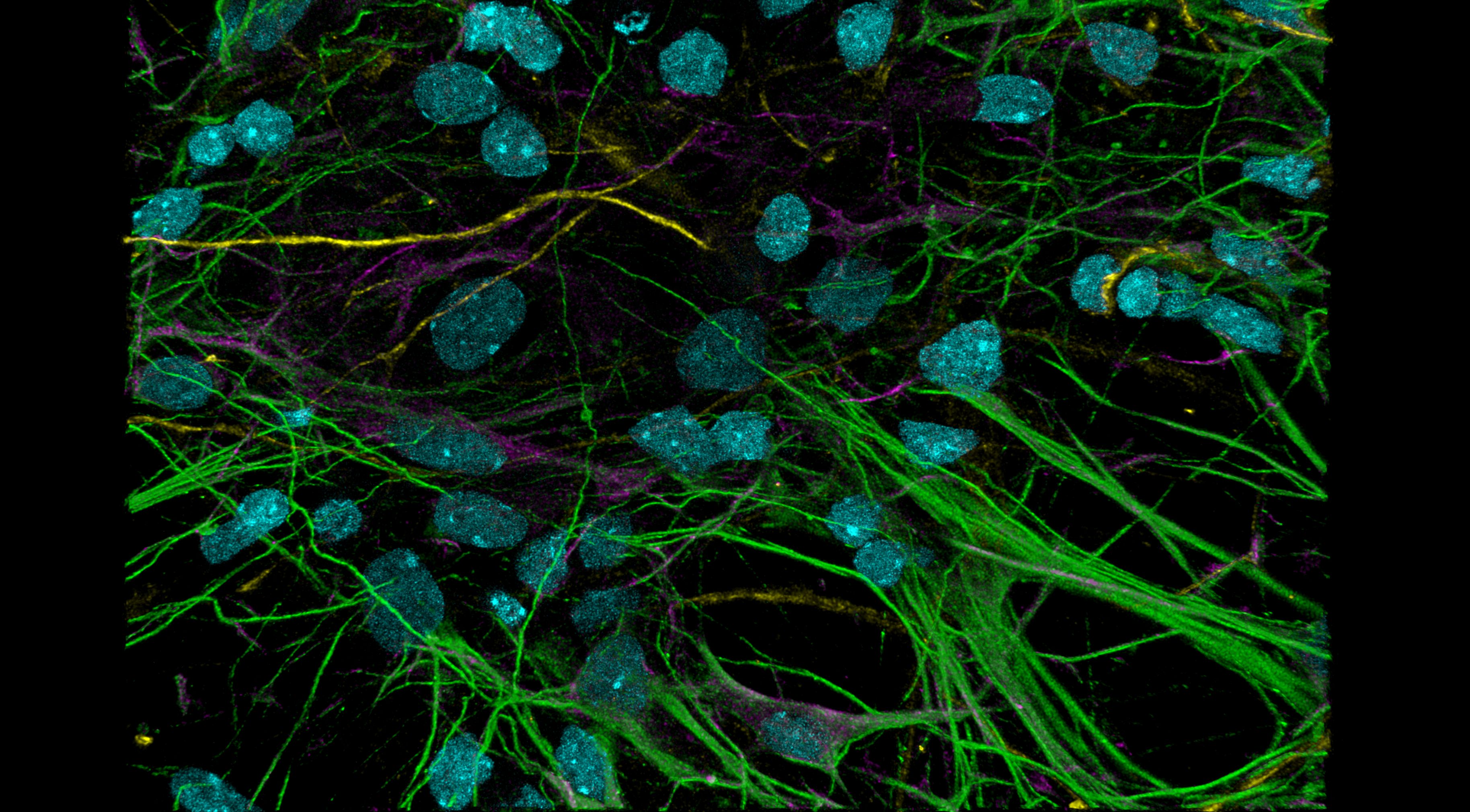
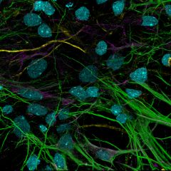 Cultured neurons stained for nuclei (Dapi, pseudo-colored in cyan) and microtubules (green). Axons are stained for the proteins nestin (yellow) and DCX (magenta). A 3D stack with 26 optical sections, 3000x3000 pixels each, was recorded with a 40x/1.4 oil objective, spanning a volume of 123x123x5 µm³. The stack was deconvolved and a projection of a 3D rendering is shown. Download high resolution image. Cultured neurons stained for nuclei (Dapi, pseudo-colored in cyan) and microtubules (green). Axons are stained for the proteins nestin (yellow) and DCX (magenta). A 3D stack with 26 optical sections, 3000x3000 pixels each, was recorded with a 40x/1.4 oil objective, spanning a volume of 123x123x5 µm³. The stack was deconvolved and a projection of a 3D rendering is shown. Download high resolution image.
Preparation: Commercial (LSM BioAnalytik). Image processing: Steffen Dietzel
|
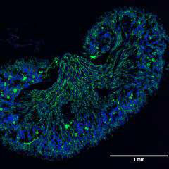 Tile scan of a neonate mouse kidney. 30 images were recorded at adjacent stage positions with 4096x4096 pixels each with the 20x/0.75 DRY objective at minimal zoom and in multi-color. Time gating was used to suppress tissue autofluorescence. Stiching created an image with 22172x18556 pixels, 4x3.34 mm and a size of 1.6 GB. The jpg image that can be downloaded here still has 39 MB. Blue: Dapi and autofluorescence. Green: endothelial cells. Red: Macrophages. Tile scan of a neonate mouse kidney. 30 images were recorded at adjacent stage positions with 4096x4096 pixels each with the 20x/0.75 DRY objective at minimal zoom and in multi-color. Time gating was used to suppress tissue autofluorescence. Stiching created an image with 22172x18556 pixels, 4x3.34 mm and a size of 1.6 GB. The jpg image that can be downloaded here still has 39 MB. Blue: Dapi and autofluorescence. Green: endothelial cells. Red: Macrophages.
Preparation: Stephan Rambichler, AG Schraml. Recording and image processing: Steffen Dietzel.
|
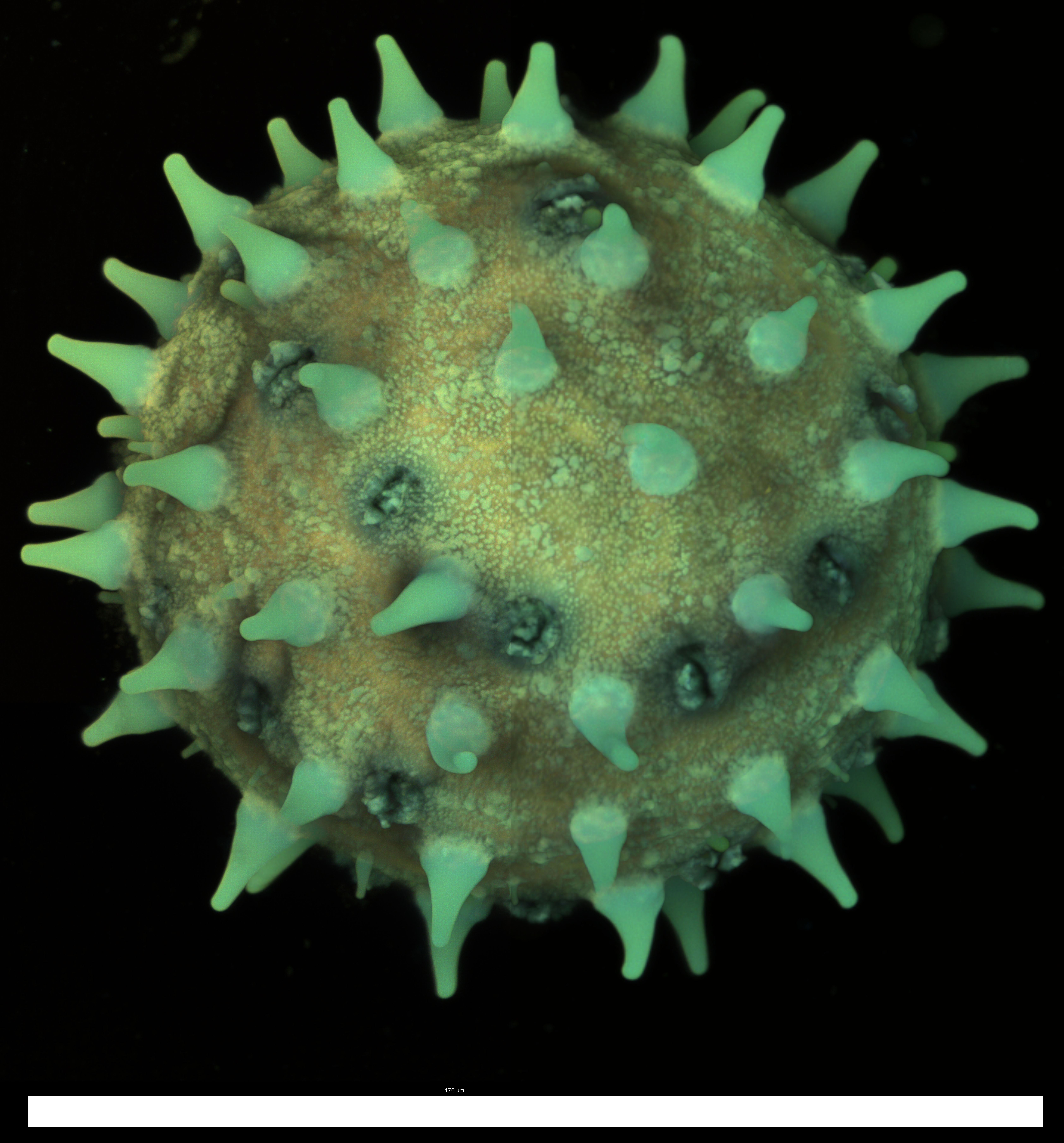
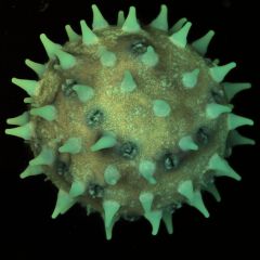 Pollen grain from Hibiscus. Maximum projection of a 3D stack with 439 optical sections and a volume of 192x192x152 µm³. Objective: 40x/1.30. Sample embedded in immersion oil. Excitation with 405 nm, autofluorescence recorded in blue, green and red. Color intensity adjusted for aesthetic impression. Download high resolution image (1.8 MB). Scale bar in high res image is 170 µm. Pollen grain from Hibiscus. Maximum projection of a 3D stack with 439 optical sections and a volume of 192x192x152 µm³. Objective: 40x/1.30. Sample embedded in immersion oil. Excitation with 405 nm, autofluorescence recorded in blue, green and red. Color intensity adjusted for aesthetic impression. Download high resolution image (1.8 MB). Scale bar in high res image is 170 µm.
Preparation, recording and image processing: Steffen Dietzel.
License: Creative Commons BY-SA 4.0
|
|
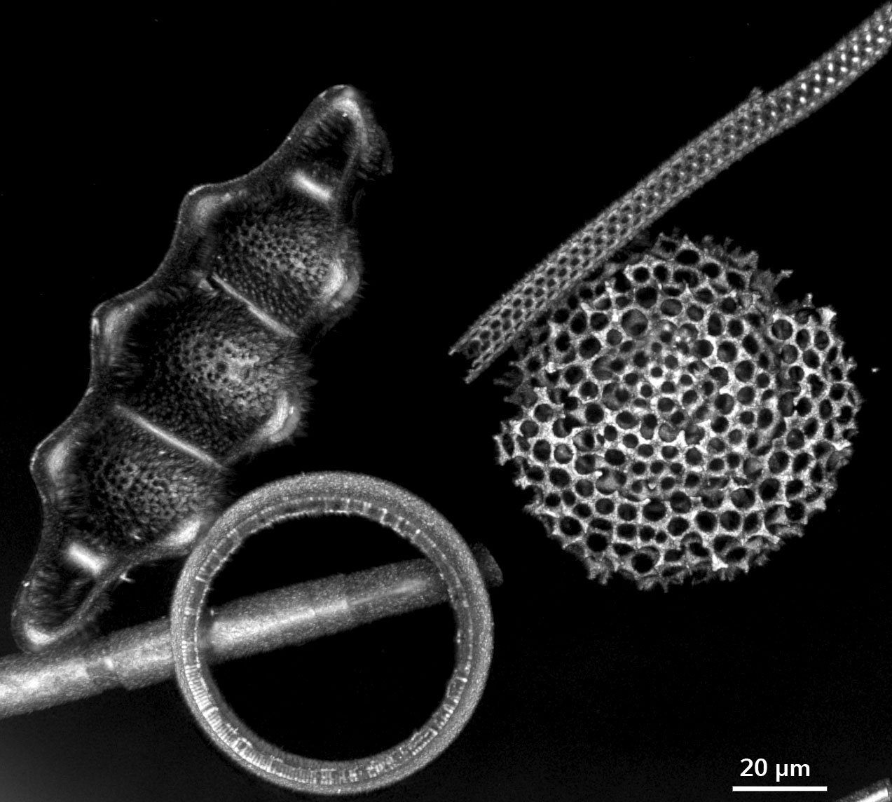
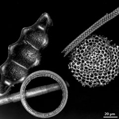 Fossile Diatoms from Oamaru (New Zealand) recorded with reflection confocal microscopy at 470 nm. On the left is a shell of Biddulphia tridens, the others are undesignated fragments. The original image stack had 129 sections with 4096x4096 pixels each, covering a volume of 234x234x33µm and was recorded with a 63x/1.40 objective. Shown is a 3D rendering of a part of the stack. Download high resolution image. Fossile Diatoms from Oamaru (New Zealand) recorded with reflection confocal microscopy at 470 nm. On the left is a shell of Biddulphia tridens, the others are undesignated fragments. The original image stack had 129 sections with 4096x4096 pixels each, covering a volume of 234x234x33µm and was recorded with a 63x/1.40 objective. Shown is a 3D rendering of a part of the stack. Download high resolution image.
Preparation: Commercial. Image recording and processing: Steffen Dietzel
|
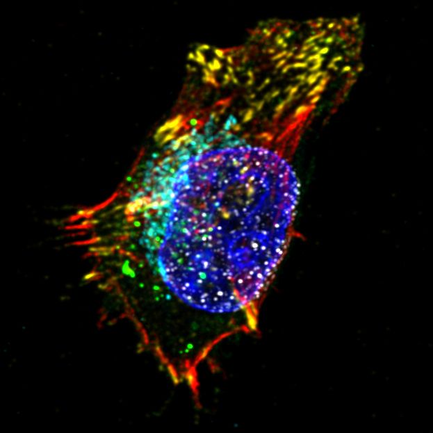
 Six color staining: HeLa cell expressing an EGFP-tagged protein that accumulates in stress granules (green). Sample was stained for nuclei (DAPI, blue), actin (Alexa Fluor 555, red), nuclear pores (Alexa Fluor 594, grey), focal adhesions (Abberior STAR 635P, yellow) and mitochondria (Alexa Fluor Plus 680, Cyan). A 3D stack with 44 optical sections, 752x752 pixels each, was recorded with a 63x/1.4 oil objective and deconvolved. Some color channels are z-projections, others are individual planes, to allow a visualization of all 6 color channels simultaneously. Download high resolution image. Six color staining: HeLa cell expressing an EGFP-tagged protein that accumulates in stress granules (green). Sample was stained for nuclei (DAPI, blue), actin (Alexa Fluor 555, red), nuclear pores (Alexa Fluor 594, grey), focal adhesions (Abberior STAR 635P, yellow) and mitochondria (Alexa Fluor Plus 680, Cyan). A 3D stack with 44 optical sections, 752x752 pixels each, was recorded with a 63x/1.4 oil objective and deconvolved. Some color channels are z-projections, others are individual planes, to allow a visualization of all 6 color channels simultaneously. Download high resolution image.
Preparation: Katarina Wachal. Image recording and processing: Andreas Thomae
|
|
|
|
|