Hooke: confocal microscope
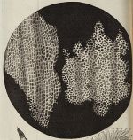 Named after Robert Hooke (1635 – 1703), an Englishman who was among many other things a famous microscopist (Wikipedia de|en). He published the best-known early book with microscopic drawings, the famous Micrographia. He also coined the term 'cell' when he noted that the microscopic structure of cork resembles the cells in a honeycomb.

Internal phone number: 71542
Internal phone directory name: Bioimaging Ost
Summary
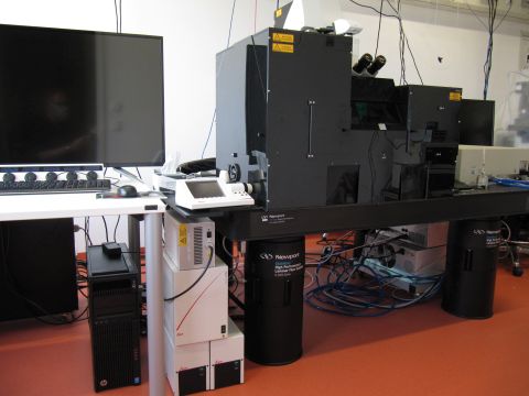
This Leica SP8 upright confocal microscope has a DM6 stand with a fixed, stable Scientifica stage. It is equipped with 405, 488, 562 and 638 nm lasers, 2 PMTs and 2 HyDs (arrangement: PMT1, PMT2, HyD3, HyD4) and transmitted light PMTs (forward external detectors; TL with 552 nm and 638 nm excitation). Multi-photon excitation is currently disabled, but would be possible continuously from 680 - 1300 nm. A CCD-Camera allows recording of conventional fluorescence. Like the microscopes Kellner, Malpighi and Gaviola, the microscope Hooke has a linear "Klondike" scanner (upgrade in 2019) resulting in a >2.5x longer dwell time (3.16 µs vs 1.2 µs) as compared to conventional sinusoidal scanners (microscope Cajal).
Introductory movie
Please go to this page for the introductory movie
Optics
Condenser
P 0.9 S1. The 0.9 indicates the numerical aperture.
Objectives
For Confocal Imaging:
- HC PL FLUOTAR 2.5x/0.07. Working distance (WD) 9.4 mm. Dry objective, with or without coverslip.
- HC PL FLUOTAR 5x/0.15. Working distance (WD) 13.7 mm. Dry objective, with or without coverslip.
- HC PL FLUOTAR 10x/0.30. Working distance (WD) 11.0 mm. Dry objective, with or without coverslip.
- HC PL APO 20x/0.75 IMM CORR CS2. Multi-immersion objective (water, glycerol, oil; adjust correction collar to your immersion medium!) for use with or without coverslip. WD 0.67 mm if used with water and 0.17 mm coverslip.
- HC PL APO 40x/1.30 Oil CS2. WD 0.24 mm. With 0.17 mm coverslip.
- HC PL APO 63x/1.40 OIL CS2. WD 0.14 mm. With 0.17 mm coverslip.
If experimental needs require another objective, please talk to the facility staff. We can borrow from other systems.
For Multiphoton Imaging (currently disabled):
HC IRAPO L 25x/1.00 W motCORR. As indicated by the name, this water immersion objective has a motorized correction collar, so that it can be adjusted to minimize spherical aberration at variable depths within the specimen, or to work with or without coverglas. Working distance is 2.6 mm. Transmission is said to be >85% between 470 and 1200 nm. "Superior" color correction from 700 to 1300 nm. This objective is mounted on its own slider (not in a revolver).
Conventional fluorescence
Meant mostly for visual inspection, but documentation is possible with the CCD-Camera. Excitation with SOLA-SM II white light LED.
Filters
The following filters for conventional fluorescence are available
- blue ("405nm LED", exc. et405/60, em et470/40, Dichroid: 455)
- green ("I3", exc. 450-490, em. LP 515)
- orange ("N2.1", exc. 515-560, em. LP 590).
CCD-Camera
Leica DFC365FX with Sony ICX285 interline CCD chip. 1392 x 1040 pixels, Chip pixel size 6.45μm. 12 bit, 4096 graylevels. Leica Website on the DFC365FX with pdf downloads.
Confocal Light Path
One-photon-Excitation Lasers
- DMOD laser 405 nm, 50 mW output, continous wave. For excitation of dyes such as DAPI.
- Solid state laser Blue, 488±2 nm, 20 mW output, continous wave.
- Solid state laser green, 552±2 nm, 20 mW output, continous wave.
- Solid state laser red, 638±2 nm, 30 mW output, continous wave.
Beam Splitter
"LIAchroic" beam splitters with low incident angle to improve transmission are used. The following are available:
- Neutral beam splitter, ratio 15/85.
- LIAchroic beam splitter for 405 / 488 / 552 nm excitation.
- LIAchroic beam splitter for 405 / 488 / 552 / 638 nm excitation.
(Note 405 excitation is not available on this system, see excitation lasers above.)
Depending on the selected excitation wavelengths, the settings for this filters are made automatically.
Detectors
Forward non-descanned detectors
The system contains two transmission detectors with which a non-confocal image can be recorded. Only compatible with 552 nm and 638 nm excitation.
Descanned, internal detectors in the spectral scan head
The descanned detectors for the confocal images are all behind the pinhole in the spectral scan head which allows continuous detection between 400 and 800 nm. The spectral scan head contains 2 Photomultiplier Tubes (PMTs; Hamamatsu R 9624) and two hybrid detectors (HyDs).
Multi-photon light path
Multi-photon excitation laser
InSight DS+ Single from Spectra Physics (product website). Emission tunable from 680 - 1300nm. Automatic dispersion precompensation. Output >1.2 W at 900nm, pulse width <120 fs.
Detectors
Four backward and two forward non-descanned, "external" detectors for multi-photon microscopy are available. In addtion to those dedicated multi-photon detectors, the internal detectors (see above) can be used with opened pinhole. While they will generally collect less light than the external ones, they may be favorable for very bright signals or for spectral windows which cannot be covered with filters for external detectors.
The external detectors are hybrid photodetectors. Light to the lower (HyD 1 & 2; green-blue) and upper deck (HyD 3 &4; red) is split by a main beam splitter separating at either 560 or 605 nm. Each deck protects the detectors with a blocking filter to suppress multi-photon excitation light (SP 800 for the upper deck). Within each deck a filter block contains a secondary beam splitter and two excitation filters.
Caution: The HyDs are very sensitive (45% QE at 530 nm) but also easily damaged! As explained during hands-on training, if you have an untested sample we highly recommend to start low excitation powers to get an impression of signal intensity. See "Sensors for True Confocal Scanning" (Leica Science Lab) for more infos on HyDs. If signals should be too bright, you also can use the internal PMTs in the spectral detector. If so, remember to open the pinhole to maximum. The following filter blocks are available:
| Name | Bandpass for channel 2/4 | Beam Splitter | Bandpass for channel 1/3 | which deck? |
|---|---|---|---|---|
| *AHF | HC 405/150 (blue) |
BS 488LPXR | ET 525/50 (green) |
lower |
| CFP-YFP | BP 483/32 (cyan) |
BS 505 | BP 535/30 (yellow) |
lower |
| FITC TRITC | BP 525/50 (green) |
BS 560 | BP 585/40 (orange) |
lower (only with main splitter 605) |
| *TRITC-A633 | BP 585/40 (orange) |
RSP 620 | BP 650/50 (far red) |
upper (only with main splitter 560) |
Cubes with * are the standard configuration. If you need other cubes, please contact the core facility staff. If the listed cubes do not satisfy your needs, have a look at the cubes for Göppert. We might be able to arrange an exchange.
Forward external detectors
The forward detectors are PMTs. Light selection is performed with a filter cube as specified in the following table. Each cube has an SP 680 blocking filter at the light entry to block multi-photon excitation light.
| Name | Channel 1 | Beam Splitter | Channel 2 |
|---|---|---|---|
| *AHF | AT 420/40 (blue) | BS 488LPXR | HC 488LP (green and longer) |
| FITC TRITC | BP 525/50 (green) | BS 560 | BP 585/40 (orange) |
The cube with * is the standard configuration. If you need the other cube, please contact the core facility staff.
Scanners
Two different scan modes are available. The "normal" scanner has a very large maximal field of view with up to 8192x8192 pixels and speeds between 1-1800 lines per second (Hz). With 512x512 pixels it can record up to 7 frames per second. Zoom factor can be 0.75 - 48x. Thanks to an upgrade installed in 2019, this scanner can be operated in a linear mode with a duty cycle of 65% (unidirectional). Thus more photons are collected in the same time.
The 8 kHz resonant scanner does a fixed number of 8000 lines per second, or 16000 in bidirectional mode. Zoom factor 1.25x - 48x.
Stage
The Scientifica MMBP stage of the microscope is fixed, meaning it cannot be automatically moved up and down. (The objective is moved up and down to make z-stacks.) Its advantage is that it can carry heavy loads, according to the spec sheet up to 30 kg. MMBP stands for Motorised Moveable Base Plate. It can be moved in x,y with a step size of 0.1 µm over 50 mm via the software or via external control.
For your Methods Section
If you used this microscope in your work, here is a suggestion for your methods section. You are welcome to copy it, but make sure to adapt it to the objectives, pixel size, dyes, filter settings etc. that you actually used.
"Multi-photon microscopy was performed at the Core Facility Bioimaging of the Biomedical Center with a Leica SP8 MP microscope, equipped with a pulsed InSight DS+ laser. Excitation of all dyes was with 860 nm. The following bandpass filters were used for detection: GFP: 525/50, Cy3: 585/40. Confocal images were recorded on the same machine using solid state laser excitation at 488, 552, and 638 nm. Images were acquired with a 20x1.0 objective, image pixel size was 200 nm. Multi-photon excited images were recorded with external, non-descanned hybrid photo detectors (HyDs), confocal images with internal conventional photomultiplier tubes."
You are very welcome to ask the facility staff for help to get your microscopy section right. Acknowledgement of the core facility is extremely important for us justify past funding and thus to secure future funding, so that we will be able to continue to provide good services to you.