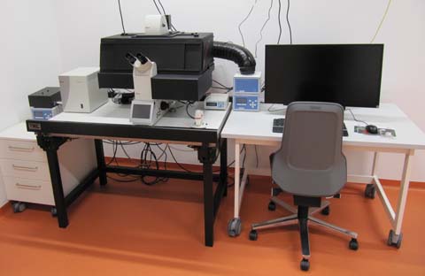
Brown: Inverted motorized live cell fluorescence microscope
 Named after Robert Brown, a British botanist who named the cell nucleus and discovered Brownian motion (Wikipedia de|en). Manufacturer's designation: Leica DMi8. Inverted fluorescence microscope with fast switchable LED excitation, motorized x,y,z movements, incubaton chamber.
Summary
This inverted Leica DMi8 is equiped for live cell imaging. Motorized stage allows time-laps recording of several postitions in parallel. Or tile scans of large areas. LED fluorescence excitation is fast switchable and avoids damaging UV light that is a problem using Mercury based lamps. Transmission white light is also LED based. Adaptive Focus Control (AFC) hardware allows long-term recording without loosing the focus. Temperature can be regulated from ambient to 37°C with the large incubator. CO2 can be added and a "water immersion mikro dispenser" for the 63x water objective allows long term observations without evaporation of immersion water.
Optics
Motorized condenser, N.A. 0.55.
Phase contrast for 10x, DIC for 63x and 100x.
Magnification Changer (1.0x / 1.6x)
objectives
- HC PL FLUOTAR 5x/0.15; WD 13,7 11506224.
- HC PL FL 10x/0.30 PH1. Working distance (WD) 11.0 mm 11506507.
- HC PL APO 20x/0.80 11506529.
- HC PL APO 40x/0.95 CORR 11506415
- HC PL APO 63x/1.40 - 0.60 oil, 11506349. Iris to reduce the NA of the objective.
- HC PL APO 100x/1.40 - 0.70 oil, 11506220. WD 0.09 mm. Iris to reduce the NA of the objective.
other objectives
- HC PL FL L 20x/0.40 CORR PH1. Long working distance (WD 6.9) obective with correction collar to adjust glass thickness from 0 - 2 mm
- HCX PL FL L 40x/0.60 CORR PH2. Long working distance (WD 3.3 - 1.9 mm) obective with correction collar to adjust glass thickness from 0 - 2 mm
- HC PL APO 63x/1.20 W CORR CS2. Water immersion objective for use with cover slip. Correction collar to adjust for cover slip thickness. An automated immersion water dispenser is available for long term experiments.
Light source
The fluorescence light source is a Spectra X from Lumencor. The data sheet can be downloaded from their web site: pdf (253 kb). It gives the exact spectral output. This LED light source can switch fast between the various channel, a property usefull in combination with the Quadband filter. It allows multi-color recordings at the maximum frame rate of the camera, without having to wait for the filter turret.
Note that the Spectra X has 6+1 channels, meaning that either the 550/15 nm or the 575/35 nm excitation band of the "green-yellow" LED (GY) can be used, but not both simultaneously. The switch between the two has to be made manually. Alternatively, one can also use the full GY emission range when removing bandpass filters. For a description of all seven possible excitations see table below.
Filters
The DMi8 has a filter turret with 6 positions. Therefore not all available filter cubes can be used at the same time. The following table shows combinations of SpectraX-emission settings with fitting filter cubes.
| Emission color | SpectraX filter | Cube name | Excitation BP | effective Excitation (both filters) | Dichroic | Emission | Typical Fluors |
|---|---|---|---|---|---|---|---|
| Blue | 390/22 | LED 405 | 405/60 | 379-401 | unknown | 470/40 | Dapi |
| Cyan | 438/29 | LED 455 | 450/50 | 425-453 | unknown | 510/40 | CFP |
| Green | 470/24 | LED 470 | 470/40 | 458-482 | unknown | 525/50 | GFP, Alexa488, FITC |
| Yellow | 510/25 | LED 505 | 494/40 | 498-522 | unknown | 535/30 | YFP |
| Orange | 550/15* | RHOD | 546/10 | 543-551 | 560 | 585/40 | Cy3, TRITC, Tomato |
| Near Red | 575/35* | LED 590 | 577/25 | 565-589 | 590 | 615/40 | TxRed, Cy3.5, Cherry, A594 |
| Far Red | 640/30 | LED 620 | 620/60 | 625-650 | unknown | 700/75 | Cy5, Draq5 |
| Quad | 390/22, 470/24, 550/15§, 640/30 | LED Sedat Quad | none | see SpectraX filter | 415, 490, 570, 660 |
none | Dapi, GFP, Cy3, Cy5 |
*Exclusive options, either 550/15 or 575/35 or no SpectraX Filter can be used. See above, description of the Spectra X light source. 575/35 and no filter is not compatible with 550 excitation and Quad detection.
A number in the form 525/50 indicates a bandpass filter with the central wavelength (525 nm) and the spectral width of the window (50 nm). In this example, the filter thus opens at 500 nm and closes at 550 nm. §Mandatory when using Quad Cube and the GY (550) excitation LED, otherwise one obtains reflection images.
Quadband filter combinations
The quad band filter cube allows fast alternating visualization of blue, green, orange and far red fluorochromes. Single fluorochrome fluorescence with the Quadband filter is achieved by choosing a single excitation LED and matching emission filter wheel position. Note that the following values are not displayed entirely correctly in the software. The following table contains the actual values (filter cube set Sedat Spectra X, k. 11525365).
| Emission color | SpectraX filter | Dichroic | Emission filter wheel |
|---|---|---|---|
| blue | 390/22 | 415 | 430/35 |
| green | 470/24 | 490 | 515/40 |
| orange | 550/15 | 570 | 595/40 |
| far red | 640/30 | 660 | 720/100 |
Camera
In 2020, the previously installed CCD-Camera was replaced with Leica DFC9000 GT sCMOS camera. While it is said to be somewhat more sensitive (up to 82% quantum efficiency at 580 nm), the major advantage is the vastly bigger chip. With about the same chip pixel size (6.5 instead of 6.45 µm) it has 2048x2048 pixels (4.2 megapixels) and is thus ~2.9 times bigger than the previous 1392 x 1040 chip of the Leica DFC365FX. Large areas of slides can thus be recorded much faster, in combination with the Navigator software package. Leica product website (de|en). DFC9000 GT-Flyer download (en) from Leica.
No additional magnification by mounting adapter. The following table shows the relevant data for the available objectives.
| Objective | Image Width | Resolution* | Nyquist** | Actual Image Pixel Size | Image Width with 1.6x Mag Lense$ | Pixel Size with 1.6x Mag Lense$ |
|---|---|---|---|---|---|---|
| 5x/0.15§ | 2.66 mm | 2033 nm | 884 nm | 1300 nm | 1.66 mm | 813 nm |
| 10x/0.30§ | 1.33 mm | 1017 nm | 442 nm | 650 nm | 832 µm | 406 nm |
| 20x/0.40 | 666 µm | 763 nm | 332 nm | 325 nm | 416 µm | 203 nm |
| 40x/0.60 | 333 µm | 508 nm | 221 nm | 163 nm | 208 µm | 102 nm |
| 20x/0.80§ | 666 µm | 381 nm | 166 nm | 325 nm | 416 µm | 203 nm |
| 40x/0.95§ | 333 µm | 321 nm | 140 nm | 163 nm | 208 µm | 102 nm |
| 63x/1.20 | 211 µm | 254 nm | 111 nm | 103 nm | 132 µm | 64 nm |
| 63x/1.40§ | 211 µm | 218 nm | 95 nm | 103 nm | 132 µm | 64 nm |
| 100x/1.40§ | 133 µm | 218 nm | 95 nm | 65 nm | 83 µm | 41 nm |
*Resolution in the focal plane (x,y) according to the Rayleigh criterion, for a wavelength of 500 nm.
** The Nyquist criterion defines the required image pixel size to realize the physically available resolution. Calculated as resolution/2.3. §Objectives currently mounted. $Imaging can be done with or without an additional 1.6x magnifying lense in beam path. The lense can be changed either at the microscope or within the LASX software: "Magnification Changer" (within the objectives/magnification menu).