Zernike: Bright field microscope with color camera
 Named after Frits Zernike (1888 - 1966), a Dutch physicist who invented phase contrast microscopy (Wikipedia de|en).
Summary
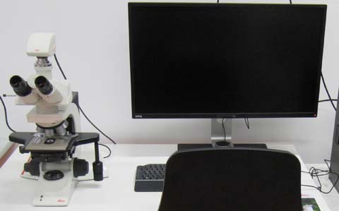
Leica DM2500. Upright microscope equipped for bright field, dark field and phase contrast. With color camera to allow recordings of classic stainings such as Hematoxylin-Eosin (HE).
Optics
Condenser UCL 0,90/1.25 Oil S1, to be used without or with oil immersion.
Objectives
HC PL FLUOTAR 5x/0.15; working distance (WD) 13.7 mm. Usable with or without coverslip.
HC PL FL 10X/0.30 PH1; WD 11.0 mm. Usable with or without coverslip.
HCX PL FLUOTAR 20x/0.50 PH2. WD 11.0mm. Usable with or without coverslip.
HCX PL FL 40X/0.75 PH2 0.17. WD 0.40mm. 0.17 mm coverslip must be used
HCX PL FLUOTAR 63x/1.25 Oil PH3. WD 0.19mm. 0.17 mm coverslip must be used
HCX PL APO 100x/1.40 Oil Ph3. 0.17 mm coverslip must be used
Camera
Leica DMC2900 CMOS Sensor camera with 2048x1536 pixel (3.1 Mpixels). Live image with 1024x768 pixels at 30 frames per second. CMOS chip pixel size 3.2 μm x 3.2 μm. The camera is mounted with a 0.55x C-mount adapter, effective pixel size is thus 5.81 µm. Example: With the 10x objective, image pixel size is 0.581 µm.
Leica Website on the DMC2900: de|en. Brochure download at Leica: de|en (pdf, 1.4 Mb)
| Objective | c-mount | Image Pixel Size |
|---|---|---|
| 5x | 0.55x | 1.162 µm |
| 10x | 0.55x | 0.581 µm = 581 nm |
| 20x | 0.55x | 0.291 µm = 291 nm |
| 40x | 0.55x | 0.145 µm = 145 nm |
| 63x | 0.55x | 0.092 µm = 92 nm |
| 100x | 0.55x | 0.058 µm = 58 nm |
For your Methods Section
If you used this microscope in your work, here is a suggestion for your methods section. You are welcome to copy it, but make sure to adapt it to the objectives, pixel size, and modality (phase contrast, bright field or dark field) that you actually used.
"Microscopy was performed at the core facility bioimaging of the Biomedical Center with a Leica DM2500 microscope equipped with a DMC2900 CMOS camera. A HCX PL FLUOTAR 63x/1.25 Oil was used for phase contrast, resulting in an image pixel size of 92 nm."
You are very welcome to ask the facility staff for help to get your microscopy section right. Acknowledgement of the core facility is extremely important for us justify past funding and thus to secure future funding, so that we will be able to continue to provide good services to you.
Quality control
Measurements of PSFs, chromatic aberration etc. will be published here as they become available.