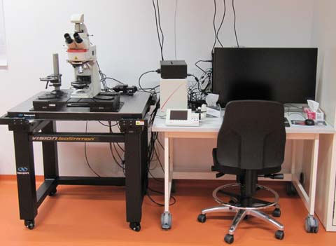
Harvey: Upright motorized fluorescence microscope
 Named after William Harvey (1578–1657), an English physician who was the first to describe that blood circulates through the human body (Wikipedia de|en).
" ...in wasps, hornets and flies even, with the aid of a magnifiying glass made to reveal the smallest things, I have seen the heart beating ... and I have shown it to others."
William Harvey in 'De motu cordis', 1578, about a quarter
century before the first microscopes were manufactured.
Translation from Latin after: An anatomical disputation concerning the movement of the heart and blood in living creatures, Gweneth Whitteridge, 1976, Blackwell Scientific Publications, Oxford, UK.
Summary
This Leica DM6 FS (Fixed Stage) fluorescence microscope can be used with all preparations requiring an upright stand, including standard microscopic slides. The stable, motorized stage allows tile scanning (overlapping images combined in one big image) and mosaic scanning (several different locations over time). LED fluorescence excitation avoids damaging UV light that is a problem using Mercury based lamps. Transmission white light is also LED based. Two cameras are available, a sensitive b/w camera for fluorescence and a color camera for bright field imaging, e.g. for HE stained slides.
Optics
Condenser N.A. 0.9.
DIC available for 100x.
Objectives
Standard objective turret (please note: objectives have higher NA and thus higher resolution and light efficiency than typical objectives of the same magnification):
- HC PL FLUOTAR 5x/0.15. Working distance (WD) 13.7 mm. Dry objective, with or without coverslip.
- HC PL FLUOTAR 10x/0.32 (11506521). WD 11.13 mm. Dry objective, best with 0.17 mm coverslip.
- HC PL APO 20x/0.80 (11506529). WD 0.4 mm. Dry objective, with 0.17 mm coverslip.
- HC PL APO 40x/0.95 CORR (11506414). WD 0.17 mm. Dry objective with 0.17 mm coverslips. Correction possible from 0.11 - 0.23 mm. The maximal NA of 0.95 for an air objective gives the highest resolution possible without immersion.
- HC PL APO 63x/1.40-0.60 OIL. Oil immersion with 0.17 mm coverslips.
- HC PL APO 100x/1.40-0.70 OIL, WD 0.09 mm. Oil immersion with 0.17 mm coverslips.
- HC APO L 10x/0.30 W UVI, WD 3.6 mm. Water immersion dipping lens, without coverslip.
- HCX APO L 40x/0.80 W UVI, WD 3.3 mm. Water immersion dipping lens, without coverslip.
- HCX APO L 63x/0.90 W UVI, WD 2.2 mm. Water immersion dipping lens, without coverslip.
The 20x/1.0W has a 32 mm screw thread and must be used with a single objective slider (does not fit in the turret). It can be exchanged for the standard turret only by Core Facility staff.
- HCX APO L20x/1.00 W. Working distance 2 mm. Water immersion dipping lens, without coverslip. If you need this objective please contact the core facility staff so that we can exchange it against the standard revolver of objectives described below. Please do not do this yourself.
Light source
The fluorescence light source is a Spectra X from Lumencor. The data sheet can be downloaded from their web site: pdf (253 kb). It gives the exact spectral output. The Spectra X has 6+1 channels, meaning that the 550 or the 575 nm excitation can be used, but not both simultaneously. The switch between the two has to be made manually. The excitation to be used with the quad band filter can be selected by fast filter wheels.
Filters
The DM6 has a fluorescence filter turret with 5 positions. One is left empty for bright field color images. Therefore only four fluorescence filter cubes can be used at the same time.
Single fluorochrome filter cubes
Single fluorochrome filter cubes provide best color visualization (highest intensity, minimal bleed through) of the fitting fluorochromes. However, switching between different fluorochromes is relatively slow. When using single fluorochrome filter cubes, excitation and emission filter wheels are set to 100% transmission.
The following table shows SpectraX filters and the properties of the respective filter cubes. For space restrictions, only 3 cubes can be mounted at the same time (one slot kept for the Quadband cube, see below). Not for all emissions of the SpectraX a fitting single fluorochrome filter cube is available.
| Emission color | SpectraX filter | Cube name | Excitation BP | effective Excitation (both filters) | Dichroic | Emission | Typical Fluors |
|---|---|---|---|---|---|---|---|
| Blue | 390/22 | ||||||
| Cyan | 438/29 | ||||||
| Green | 470/24 | GFP-ET | 450-490 | 458-482 | 495 | 500-550 | GFP, Alexa488, FITC |
| Yellow | 510/25 | ||||||
| Orange* | 550/15 | Cy3 | 541-551 | 543-551 | 556 | 560-584 | Cy3, TRITC, Tomato |
| Near Red* | 575/35 | Cy3.5 | 562-592 | 562-592 | 590 | 610-640 | TxRed, Cy3.5, Cherry, A594 |
| Far Red | 640/30 | Y5-ET | 590-650 | 625-650 | 660 | 662-738 | Cy5, Draq5 |
*Exclusive options, either orange or near red can be used. See above, description of the Spectra X light source. The standard setting is the orange filter. The cube for Cy3.5 has to be set up by facility personnel.
A number in the form 550/15 indicates a bandpass filter with the central wavelength (550 nm) and the spectral width of the window (15 nm).
Quadband filter combinations (as of 10/2020)
The quad band filter cube allows fast alternating visualization of blue, green, orange and far red fluorochromes. Single fluorochrome fluorescence with the Quadband filter is achieved by an excitation and emission filter wheel which can switch filters very fast. Note that the following values are not displayed entirely correctly in the software. The following table contains the actual values (filter cube set DFT51111, k. 11504258).
| Emission color | SpectraX filter | Excitation filter wheel | Excitation band in Quadband | Dichroic | Emission band in Quadband | Emission filter wheel |
|---|---|---|---|---|---|---|
| blue | 390/22 | 395/25 | 391/32 | 415 | 435/30 | 440/40 |
| green | 470/24 | 480/30 | 479/33 | 500 | 519/25 | 520/40 |
| orange | 550/15 | 555/25 | 554/24 | 572 | 594/32 | 590/50 |
| far red | 640/30 | 640/30 | 638/31 | 660 | 695/58 | 700/75 |
Cameras
B/w camera for fluorescence
In 2020, the previously installed CCD-Camera was replaced with Leica DFC9000 GT sCMOS camera. While it is said to be somewhat more sensitive (up to 82% quantum efficiency at 580 nm), the major advantage is the vastly bigger chip. With about the same chip pixel size (6.5 instead of 6.45 µm) it has 2048x2048 pixels (4.2 megapixels) and is thus ~2.9 times bigger than the previous 1392 x 1040 chip of the Leica DFC365FX. Large areas of slides can thus be recorded much faster, in combination with the Navigator software package. Leica product website (de|en). DFC9000 GT-Flyer download (en) from Leica.
No additional magnification by mounting adapter. The following table shows the relevant data for the available objectives.
| Objective | Resolution* | Nyquist** | Actual Image Pixel Size | Image Width |
|---|---|---|---|---|
| 5x/0.15 | 2033 nm | 884 nm | 1300 nm | 2.66 mm |
| 10x/0.32 | 953 nm | 414 nm | 650 nm | 1.33 mm |
| 20x/0.8 | 381 nm | 166 nm | 325 nm | 666 µm |
| 40x/0.95 | 321 µm | 140 nm | 163 nm | 333 µm |
| 63x/1.40 oil | 218 nm | 95 nm | 103 nm | 211 µm |
| 100x/1.40 oil | 218 nm | 95 nm | 65 nm | 133 µm |
| water imm: | ||||
| 10x/0.3 | 1017 nm | 442 nm | 650 nm | 1.33 mm |
| 40x/0.8 | 381 nm | 166 nm | 163 nm | 333 µm |
| 63x/0.9 | 339 nm | 147 nm | 103 nm | 211 µm |
| 20x/1.0 | 305 nm | 133 nm | 325 nm | 666 µm |
*Resolution of the objective in the focal plane (x,y) according to the Rayleigh criterion, for a wavelength of 500 nm.
** The Nyquist criterion defines the required image pixel size to realize the physically available resolution. Calculated as resolution/2.3.
Color Camera for bright field
Leica DMC2900 CMOS Sensor camera with 2048x1536 pixel (3.1 Mpixels). Live image with 1024x768 pixels at 30 frames per second. CMOS chip pixel size 3.2 μm x 3.2 μm. The camera is mounted with a 0.55x C-mount adapter, effective pixel size is thus 5.81 µm. Example: With the 10x objective, image pixel size is 0.581 µm.
Leica Website on the DMC2900: de|en. Brochure download at Leica: de|en (pdf, 1.4 Mb)
| Objective | Resolution* | Nyquist** | Image Pixel Size |
|---|---|---|---|
| 5x/0.15 | 2 µm | 884 nm | 1.162 µm |
| 10x/0.30 | 1 µm | 442 nm | 581 nm |
| 20x/1.0 | 0.3 µm | 133 nm | 291 nm |
| 40x/0.8 | 0.38 µm | 166 nm | 145 nm |
| 63x/0.9 | 0.34 µm | 147 nm | 92 nm |
| 63x/1.40 | 0.22 µm | 95 nm | 92 nm |
| 100x/1.40 | 0.22 µm | 95 nm | 58 nm |
*Resolution in the focal plane (x,y) according to the Rayleigh criterion, for a wavelength of 500 nm.
** The Nyquist criterion defines the required image pixel size to realize the physically available resolution. Calculated as resolution/2.3.
For your Methods Section
If you used this microscope in your work, here is a suggestion for your methods section. You are welcome to copy it, but make sure to adapt it to the objectives, camera, image pixel size, and modality (fluorescence, bright field) that you actually used.
"Microscopy was performed at the Core Facility Bioimaging of the Biomedical Center with a Leica DM6 FS microscope. A 40x/0.95 objective was used. Fluorescence was recorded with a Leica DFC9000 GT sCMOSL camera (note: up to 09/2020 Leica DFC365FX camera) with an image pixel size of 163 nm. Bright field images were recorded with a Leica DMC2900 CMOS camera with an image pixel size of 145 nm."
You are very welcome to ask the facility staff for help to get your microscopy section right. Acknowledgement of the core facility is extremely important for us justify past funding and thus to secure future funding, so that we will be able to continue to provide good services to you.