Stokes: Fluorescence stereo microscope
 Named after Sir George Gabriel Stokes (1819 - 1903), an Irish mathematician and physicist who discovered and named fluorescence (Wikipedia de|en).
Summary
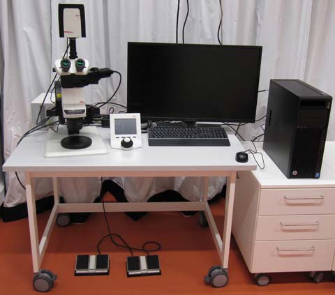
Leica M205 FA stereo microscope. Large enough working distance to be used for preparations. Two foot paddles to adjust magnification and focus. Magnification up to over 200x, camera for documentation. Incident white light illumination as well as fluorescence excitation and filters for blue, green and orange/red channels.
A somewhat different light path for the left and right eye combines a large depth of focus with good z-discrimination. z-stacks can be recorded.
Optics
This stereo microscope comes with two objectives. The Planapo 1x is the standard objective for most application. The Planapo 2x can be used where higher magnification is needed. The exchange must be made by manually unscrewing the one and screw in the other objective. Due to the weight of the objectives this bears the risk of damage. Therefore, please contact the facility staff if you need the 2x objective.
The eyepieces (10x/23B) magnify ten times. The remaining magnification is generated by the internal zoom optics. With the eye pieces, total zoom ranges from 7.82x to 160x
A circle of 29.5 mm can be observed with the eye pieces at minimal zoom (7.82x). The camera image at minimal zoom has a size of 14 mm by 10.5 mm.
Fluorescence
Light source
Lumencor SOLA-SM-II white LED.
Filters
Filter system ET DAPI BP: Excitation BP 383 - 407 nm; emission BP 435 - 485 nm. (Blue fluorochromes, e.g. DAPI)
Filter system ET GFP: Exc. BP 450 - 490 nm; em. BP 500 - 550 nm. (Green fluorochromes, e.g. GFP)
Filter system ET dsRED: Exc. BP 530 - 560; em. BP 590 - 650 nm. (Red fluorochromes, e.g. dsRed)
Since fluorescence excitation and emission run through a slightly different beam path, no dichroics are used.
Camera
Leica DFC7000 T
For your Methods Section
If you used this microscope in your work, here is a suggestion for you method section. You are welcome to copy it, but make sure to adapt it to the objectives, dyes and filters that you actually used.
"Fluorescence microscopy was performed at the core facility bioimaging of the Biomedical Center with a Leica M205 FA stereo microscope, equipped with a DFC7000 T camera. Images were acquired with a Planapo 1x objective, zoom was set to XX1, resulting in an image pixel size of XX1 µm. The following fluorescence channels were used: blue (exc 395/25 nm; em 460/50 nm), green (470/40; 525/50), red (545/30; 620/60)."
1 This information can be found in the Leica image file. If you do not find it, the facility staff can show you how.
You are very welcome to ask the facility staff for help to get you microscopy section right. Acknowledgement of the core facility is extremely important for us to secure future funding and thus to provide good services to you.