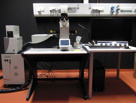
Cajal: Confocal Microscope (NC00.058)
 Named after Santiago Ramón y Cajal (1852 -1934), a Spanish pathologist, neuroscientist, and Nobel prize winner who became famous for his drawings of the nervous system (Wikipedia de|en). He also wrote a book "Advice for a Young Investigator" (Reglas y Consejos sobre Investigación Cientifica: Los tónicos de la voluntad) which can be found as pdf on the Internet.
Summary
This "Leica TCS SP8 with compact supply unit" has an inverted stand (DMi8). It is equipped with four laser lines (405, 488, 552 and 638 nm), one PMT and three hybrid detectors.
Optics
Long distance condenser with condenser head S28/0.55
Objectives
- HCPL FLUOTAR 10x/0.30, working distance (WD) 11.0 mm. Usable with and without coverslip.
- HC PL APO 20x/0.75 IMM CORR CS2. Usable with and without coverslip, with water, glycerol or oil. WD depends on immersion, e.g. 0.67 mm for water with coverslip.
- HC PL APO 40x/1.30 Oil CS2. WD 0.24 mm.
- HC PL APO 63x/1.40 OIL PH3 CS2. With phase contrast ring, WD 0.14 mm.
- HC PL APO 100x/1.40 OIL CS2. WD 0.13 mm.
Conventional fluorescence
Meant for visual inspection only, not for documentation. Excitation with EL6000(LQHXP 120 LEJ). Filters for blue (exc. 325-375, em. LP 435-485), green (exc. 460-500, em. 512-542) and orange (exc. 541-551, em. 565-605) are available.
Confocal Light Path
Excitation Lasers
Four lasers are available, each providing one line.
- 405 nm, 50 mW. For excitation of dyes such as DAPI.
- 488±2 nm, 20 mW. For excitation of dyes such as GFP, Alexa488, FITC.
- 552±2 nm, 20 mW. For excitation of dyes such as Cy3, TRITC.
- 638±2 nm, 30 mW. For excitation of dyes such as Cy5, AberriorStar635P, SiR.
Each laser can be attenuated individually to the required intensity.
Beam Splitters
The system works with LIAchroics low incident angle dichroic beam splitters. The following can be used:
- For 405 / 488 / 552
- For 405 / 488 / 552 / 638
- Neutral splitter with a 15/85 ratio
Detectors
The system contains one transmission detector with which a non-confocal image can be recorded.
The detectors for the confocal images are all in the spectral scan head which allows continous detection between 400 and 800 nm. We have 2 Photomultiplier Tubes (PMT; Hamamatsu R 9624)) and 3 hybrid photo detectors (HyDs). Caution: The HyDs are more sensitve (45% QE at 530 nm) but also more easily damaged! As explained during hands-on training, if you have an untested sample we highly recommend to start with the PMT to get an impression of signal intensity. See "Sensors for True Confocal Scanning" (Leica Science Lab) for more infos on HyDs.
Scanners
Two different scan modes are available. The "normal" scanner has a very large maximal field of view with up to 8192x8192 pixels and speeds between 1-1800 lines per second (Hz). With 512x512 pixels it can record up to 7 frames per second. Zoom factor can be 0.75 - 48x.
The 8 kHz resonant scanner does a fixed number of 8000 lines per second, or 16000 in bidirectional mode. It records 28 frames at 512x512 pixels. Maximal image size is 1248x1248 pixels, zoom factor 1,3x - 48x.
For your Methods Section
If you used this microscope in your work, here is a suggestion for your methods section. You are welcome to copy it, but make sure to adapt it to the objectives, pixel size, dyes, filter settings etc. that you actually used.
"Confocal microscopy was performed at the core facility bioimaging of the Biomedical Center with an inverted Leica SP8 microscope, equipped with lasers for 405, 488, 552 and 638 nm excitation. Images were acquired with a 100x1.4 objective, image pixel size was 80 nm. The following fluorescence settings were used for detection: Dapi: 430-470 nm, GFP: 500-550, Cy3: 560-600, Cy5:650-700). Recording was sequentially to avoid bleed-through. GFP, Cy3 and Cy5 were recorded with hybrid photo detectors (HyDs), Dapi with a conventional photomultiplier tube."
You are very welcome to ask the facility staff for help to get your microscopy section right. Acknowledgement of the core facility is extremely important for us justify past funding and thus to secure future funding, so that we will be able to continue to provide good services to you.