Blog Entry: Immersion Oils and Chromatic Aberration
A comparision of the 'original' Leica/Zeiss immersion oil with Cargille HF with our 100x1.4 oil STED White objective
23.07.2018
High resolution microscopy needs high quality immersion oil and that is expensive. You may have seen that we have several Leica microscopes at the Core Facility Bioimaging. As they always do, they came with a starter supply of 10 ml bottles with immersion oil "Type F" (F standing for fluorescence microscopy, meaning the oil is non-fluorescent). When I first checked out their material safety data sheet I was in for a surprise: The manufacturer of this oil is Carl Zeiss. Oil is probably the only Zeiss product that is sold by Leica Microsystems. So I figured I can as well buy the Zeiss oil which is called Immersol 518F and which is available in larger bottles.
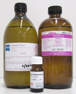 When looking around I found a supplier that would deliver a 500 ml bottle for € 515 (plus tax). That gave quite a saving compared to the same amount in 10 or 20 ml bottles but is still not exactly cheap. So I was looking for alternatives. Colleagues mentioned good experiences with Cargille Type HF. The Cargille web site currently list the 16 fl.oz. bottle (= 473 ml) for US$ 65 (~€ 55). Even with the about 200 Euro shipping costs to Germany, this would still be a bargain.
On paper, both oils are very similar. The 518 in the name of the Immersol apparently comes from the refractive index of 1.518, while the Cargille HF is announced with an Ri of 1.515. But when looking closer it turns out that these Ris are measured at different wavelengths. Both oils have an ne (@546 nm) of 1.518 and an nD (@589 nm) of 1.515, all at 23°C and according to the manufacturers. So I ordered.
What I noticed only much later is the different dispersion value, which is given as ve=46 on the Leica bottle, ve=45 on the Zeiss bottle and ve=43.6 for the Cargille oil.
The Cargille HF arrived and we did some tests, like looking at the point spread function of gold beads in reflection mode. Full width half maxima with a 63x1.4 objective on a confocal were in the same range for both oils, so everything seemed to be fine.
Only much later did we notice something odd. On our STED microscope, when doing an xz-scan on gold beads in reflection mode, the depletion donut at 775 nm seemed to be quite a bit away from the focus of the excitation beam. Reason enough to look closer. So we tested our Leica 100x1.4 STED white objective, which is advertised to have less than 50 nm chromatic aberration in z (see this Leica web page for a diagram with the calculated chromatic aberration of this and other STED objectives), with both oils and gold beads in reflection mode.
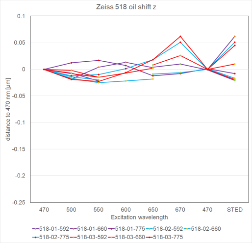
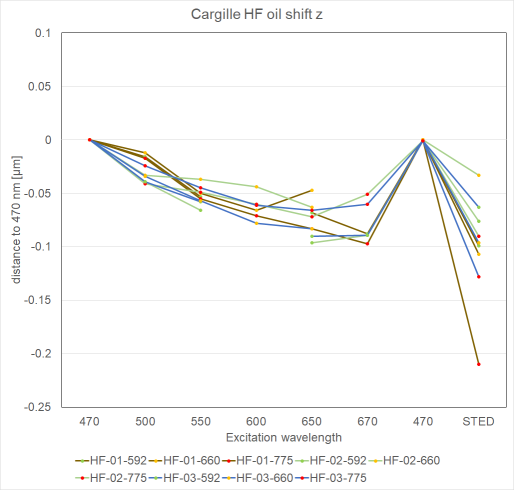
Indeed, it turned out that the chromatic aberration in z was less with the 518F oil. Using 470 nm as our reference line, at 670 nm we found up to around 50 nm chromatic aberration with 518F and up to around 100 nm with HF. (see diagrams below for details. However, when repeating measurements with the very same beads (same line color in diagram) and the same wavelength, we also found differences of up to around 50 nm in some instances. So this seems to be the error range we have to deal with for this kind of measurement. Still, comparing the two sets of measurements at large, the difference between the two oils becomes clear.
Of special interest to us were the aberrations for the 775 nm STED depletion laser (red dots at 'STED' excitation in diagram). For the 518F, all three values were at 50 nm or less, compared to 470 nm. For HF the three values were higher, at 60, 130 and 210 nm.
I do not consider the results described here a scientific study; for this I would want to have more data, given the variation of repeated experiments. But the trend already becomes quite clear, we see more chromatic aberration with HF than with 518F oil with this objective.
So, is HF a bad oil? I don't think so. 100 nm chromatic aberration in z between 470 and 670 nm wavelengths is still pretty low, the difference between the two oils is even less. For comparison, this is less than the expected chromatic aberration for a 'normal' 100x1.4 objective (~250 nm according to the Leica diagram mentioned above). Assuming that the z-resolution of a fluorescence or confocal microscope is in the range of half a micron, again the difference between the two oils is not that big.
Still, for STED, where it is important that the depletion donut is positioned exactly around the excitation spot, I'd rather use the oil which the manufacturer had in mind when calculating the objective.
Obviously the results may be different for a different objective from the same or another manufacturer. There is no substiture for testing a given setup, in particular if colocalization studies are performed.
Diagrams and further details
Chromatic aberration in z-direction with Zeiss 518F or Cargille HF, compared to the focus of 470 nm light.
Images were recorded line sequential with several wavelengths from 470 to 670 nm, again 470 nm and finally a STED depletion wavelength (see x-axis of the diagrams). Only one STED depletion line can be used in a line sequential setting. Therefore, a group of beads was recorded three times (same line color), once with each of our three depletion laser wavelengths, 592 (green dots), 660 (orange dots) and 775 nm (red dots). For the triplicate images from 470 - 670 nm this means that the same beads in theory should give the exact same result (but they don't, due to measurement errors, e.g overexposed beads in some images that were discarded).
In settings where the 592 nm depletion line was recorded, no image could be made at 600 nm, with the 660 nm depletion line no image could be made at 670 nm, due to notch filters in the beam path. Hence the gaps in the respective lines.
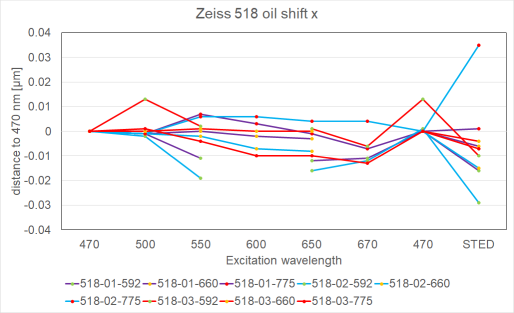
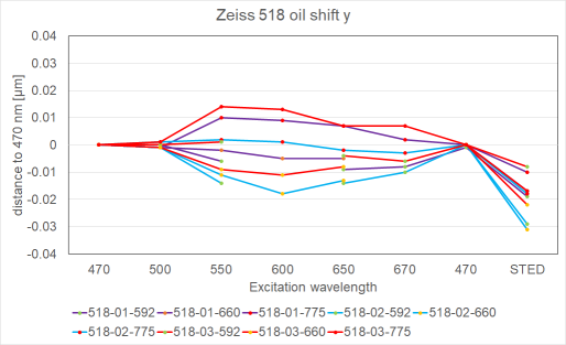
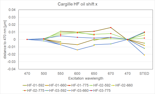
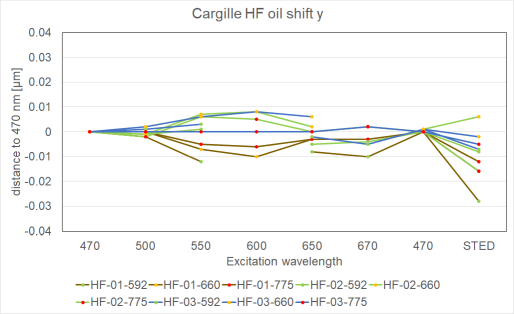
Chromatic aberrations in x,y
Chromatic aberration within the focal plane was generally less than 20 nm and thus negligible for practical purposes. Somewhat larger values for the depletion beams may be due to non-perfect alignment of the two beams or due to non-symetrical appearance of the donuts.
Methods
Images were recorded on our Leica SP8 WLL STED 3X in sequential mode with the 100x STED white objective. 80 nm gold beads embedded in ProLong Diamond were imaged in reflection mode with PMTs. Voxel size was set to 81x81x100 nm³. Images were opened in Fiji and the chromatic aberration between intenisty gravity centers was determined with the Sync Measure 3D tool which is under the Analyze>Tools menu. Diagrams were made in Excel.
Contributors
Material, data and ideas were contributed and evaluated by Katarina Wachal, Andreas Thomae and Steffen Dietzel. Text written by SD.
Any questions, comments or suggestions on this article? Please mail me.