Inauguration Symposium a great success
Over 200 participants attend the opening event
18.03.2016
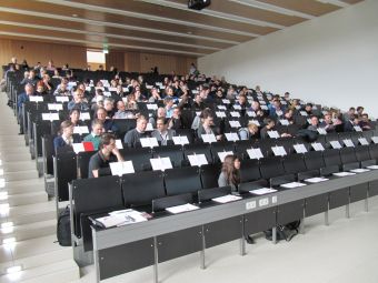 The lecture hall about 10 minutes before the start of
 the symposium, before the blinds went down
Over 180 participants registered for the Inauguration Symposium of the Core Facility Bioimaging on 17. February and even more attendees turned up on site, at the "Kleiner Hörsaal" of the BMC, the 'small lecture hall' which can hold 300 people.
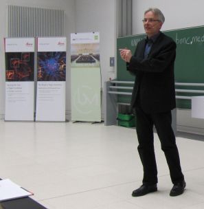
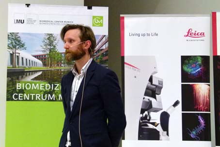
After a welcome address by Barbara Conradt, Vice President for Research of the University and chair of Cell and Developmental Biology at the neighboring Biocenter, and short introductions given by Ulrich Pohl, head of the Walter-Brendel-Zentrum für Experimentelle Medizin which hosts the Core Facility, and Steffen Dietzel, Head of the Core Facility itself, Christoph Thumser from Leica Microsystems offered his view on academic-industrial partnerships in general an on the partnership of Leica Microsystems with the Core Facility Bioimaging in particular.
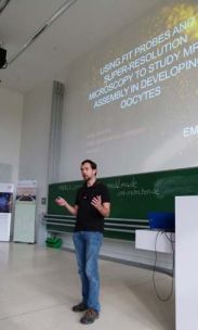
Five excellent scientific talks followed suit, covering the range from molecular level to whole organisms. Imre Gaspar from the EMBL in Heidelberg explained how to use FIT probes to perfom super-resolution microscopy for studying mRNP assembly in oocytes. FIT stands for 'forced intercalation of TO (Thiazole Orange)', a method to label nucleic acids. Elisa Greotti from the Pozzan lab at the University of Padua talked about mitochondrial signalling and how that connects physiology and pathology, e.g. in Alzheimers disease.
 Imre Gaspar and Friedemann Kiefer
After the break, Friedemann Kiefer from the Max-Planck-Institute in Münster demonstrated imaging of the vascular systems during morphogenesis and disease, with a special emphasis on the 'other' vascular system, the lymphatic vessels and how optical clearing and specific form of planar illumination can help in such analyses. William Louch from the University of Oslo discussed mechanisms of heart failure and also discussed differences between rodent and human hearts, e.g. in T-tubule numbers. Finally, Valentin Nägerl from the Université de Bordeaux gave a summery of his approaches to image the micro-anatomical mechanisms of neural plasticity, including STED microscopy in tissues.
 William Louch
After the talks, to conclude an exciting day of science and celebration, delegations from Leica and the Core Facility went to the facility rooms to perform a little ceremony, to cut a ribbon to symbolize the official opening.
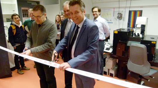 In the foreground, cutting the ribbon, Steffen Dietzel, head of the Core Facility Bioimaging and Christioph Thumser from Leica Microsystems, Head of Sales for EMEA. Between them Ulrich Pohl, head of the Walter-Brendel-Zentrum, the hosting institution of the Core Facility.