The Laser Microdissection System from Leica
Technology, Applications and Workflows
04.07.2017
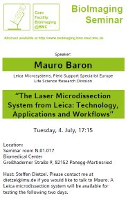
The Core Facility Bioimaging in collaboration with Leica Microsystems provides the opportunity to learn about the possibilities provided by Leica's new laser microdissection (LMD) system. Hands-on sessions with interested users are planed for the 5th and 6th July 2017. Please contact us for an appointment. The LMD system will be introduced by a talk to which all interested are invited:
"The Laser Microdissection System from Leica: Technology, Applications and Workflows"
by Mauro Baron,
Leica Microsystems, Field Support Specialist Europe Life Science Research Division
Tuesday, 4. July, 17:15
at the seminar room N.01.017 at the BMC
Abstract:
Diseases have reasons and symptoms which can be directly connected to the underlying cellular components. Researchers have the ability e.g. to search for mutations, investigate gene activity, or study the fate of gene products or their effectors. In other words, from a cellular view, diseases can be explored on the level of DNA, RNA, proteins, carbohydrates, lipids, or small molecules. To study these cellular components with the help of modern lab methods such as DNA sequencing, NGS, qPCR, or mass spectrometry their purification is recommended if not mandatory.
Laser Microdissection (LMD) is an emerging microscopic technology with increasing applications for DNA, RNA and proteomic analysis. The method is designed for contact- and contamination-free isolation of entire areas of tissue or specific single cells or sub-cellular structures, e.g. single chromosomes from a wide variety of tissue samples, allowing the isolation of homogeneous, specific and pure targets from heterogeneous samples for downstream analysis. The dissectate is then available for further molecular methods such as PCR, real-time PCR, proteomics and other analytical techniques.
LMD is now used in a large number of research fields, e.g. neurology, cancer research, plant analysis, forensics.
LMD systems offer a precise, easy and contamination-free solution to obtain sufficient sample quantities from specimens. Simply identify your region of interest drawing it on the PC screen and directly cut the areas by a guided laser beam. The dissectate is automatically separated from the surrounding tissue with the movement of a laser beam and is immediately available for downstream analysis. The whole process can be fully automated in combination with pattern recognition (e.g. AVC).
Downloads
- Mauro Baron (197 KByte)


 Download pdf-flyer


